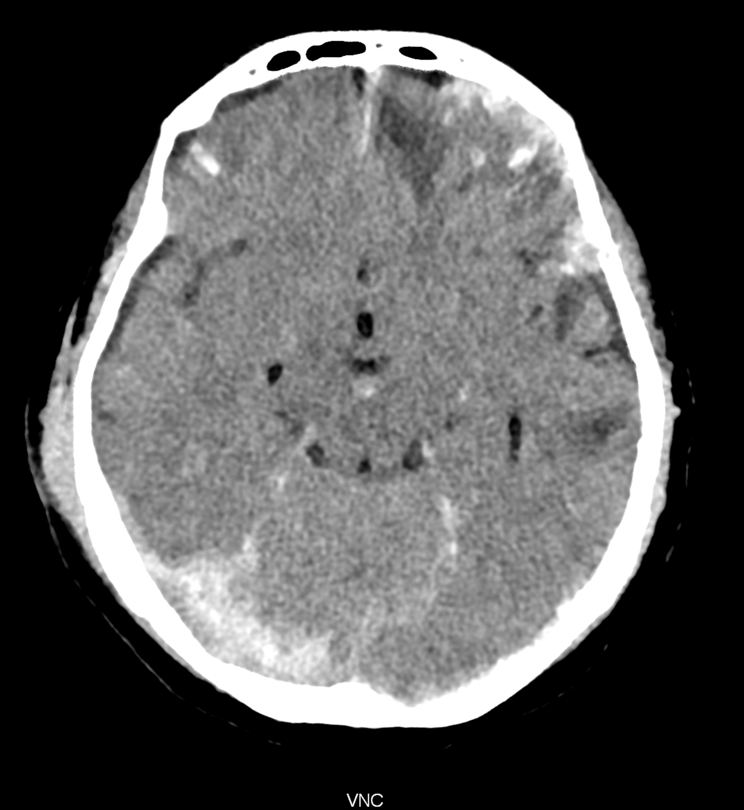
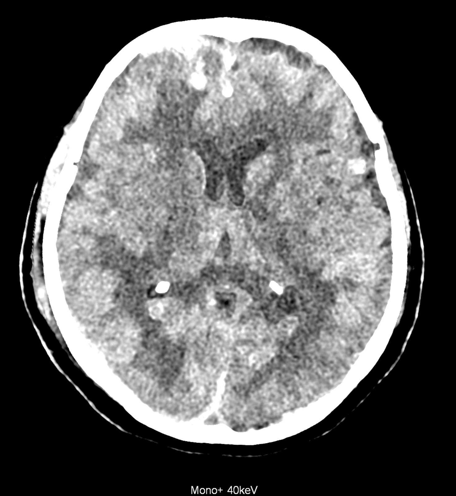
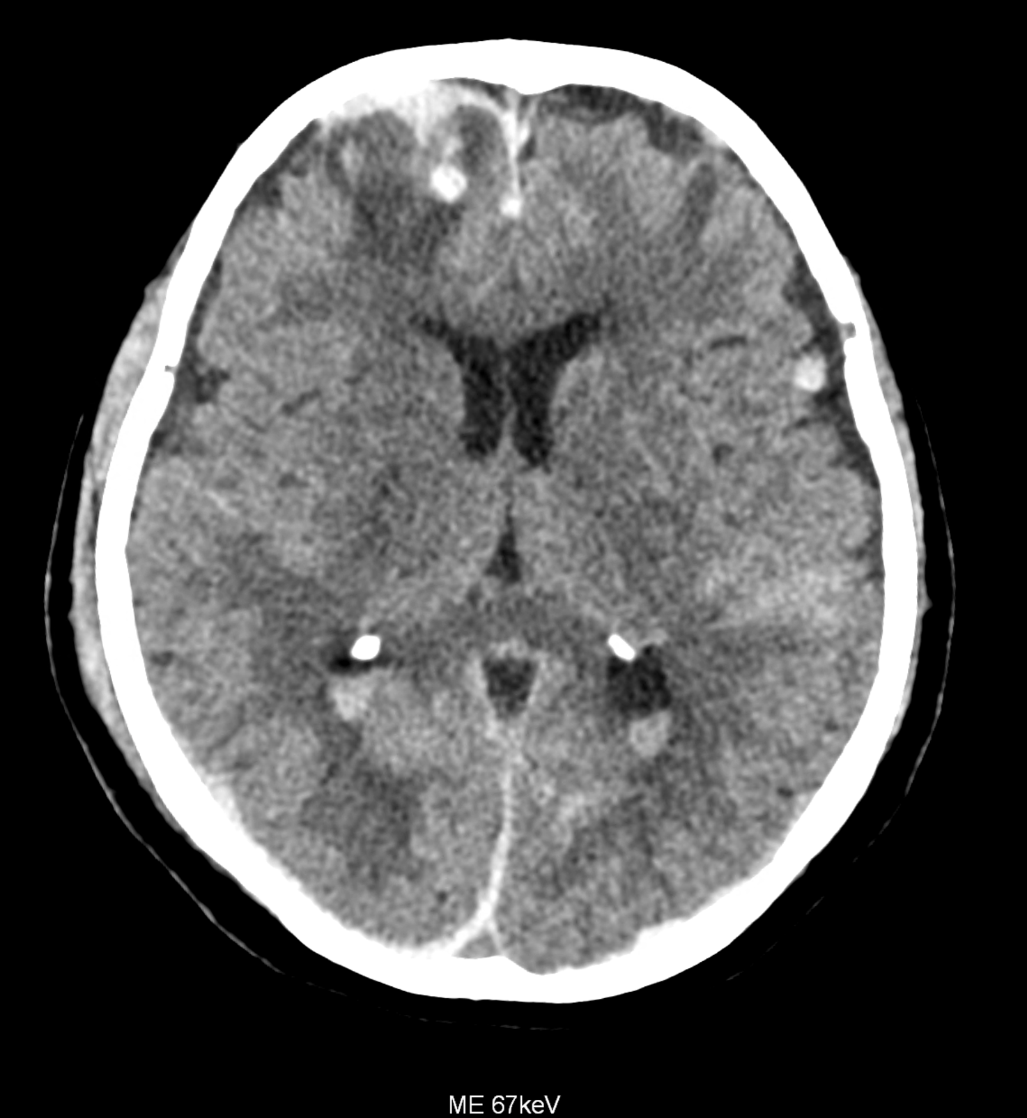
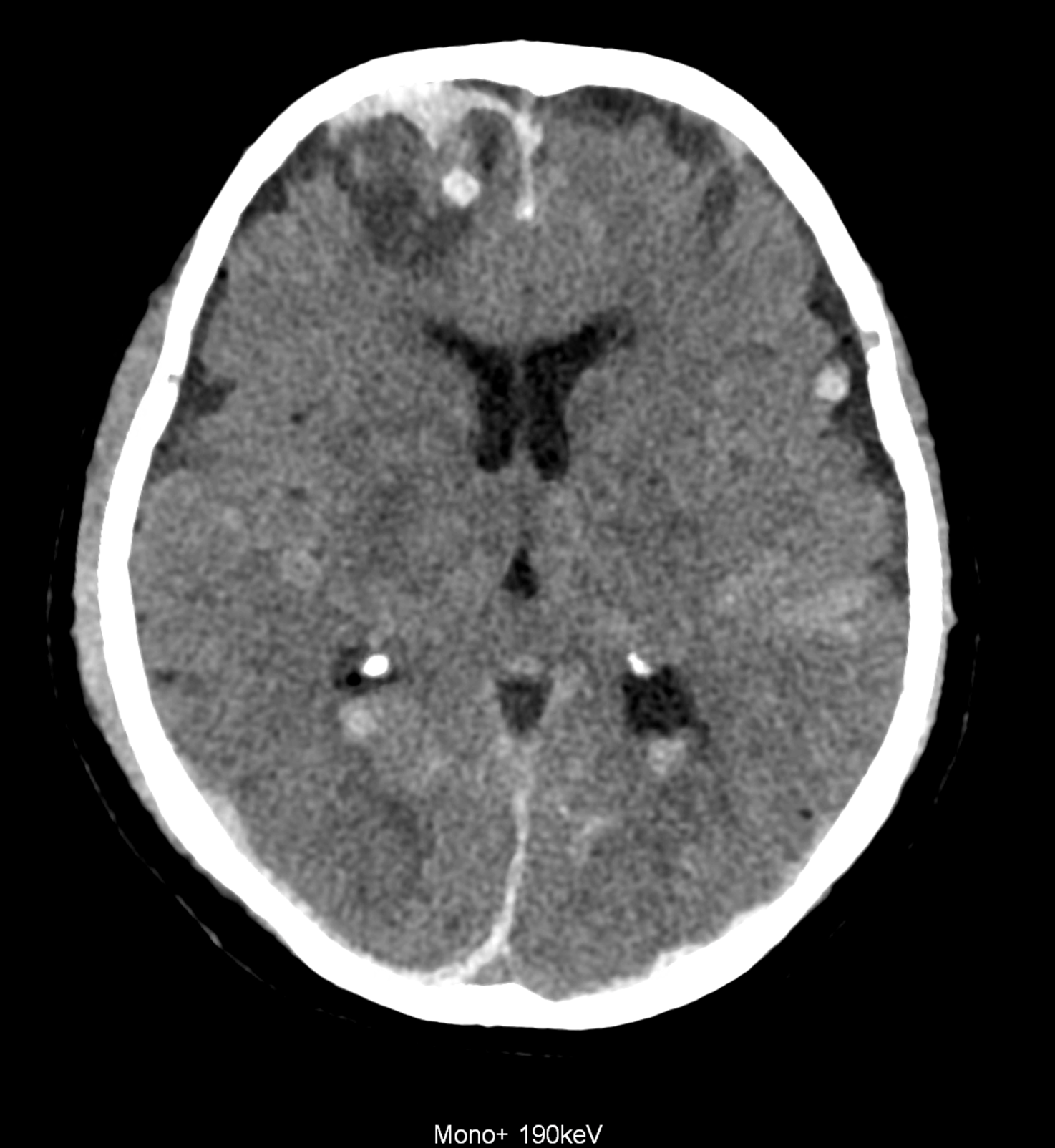
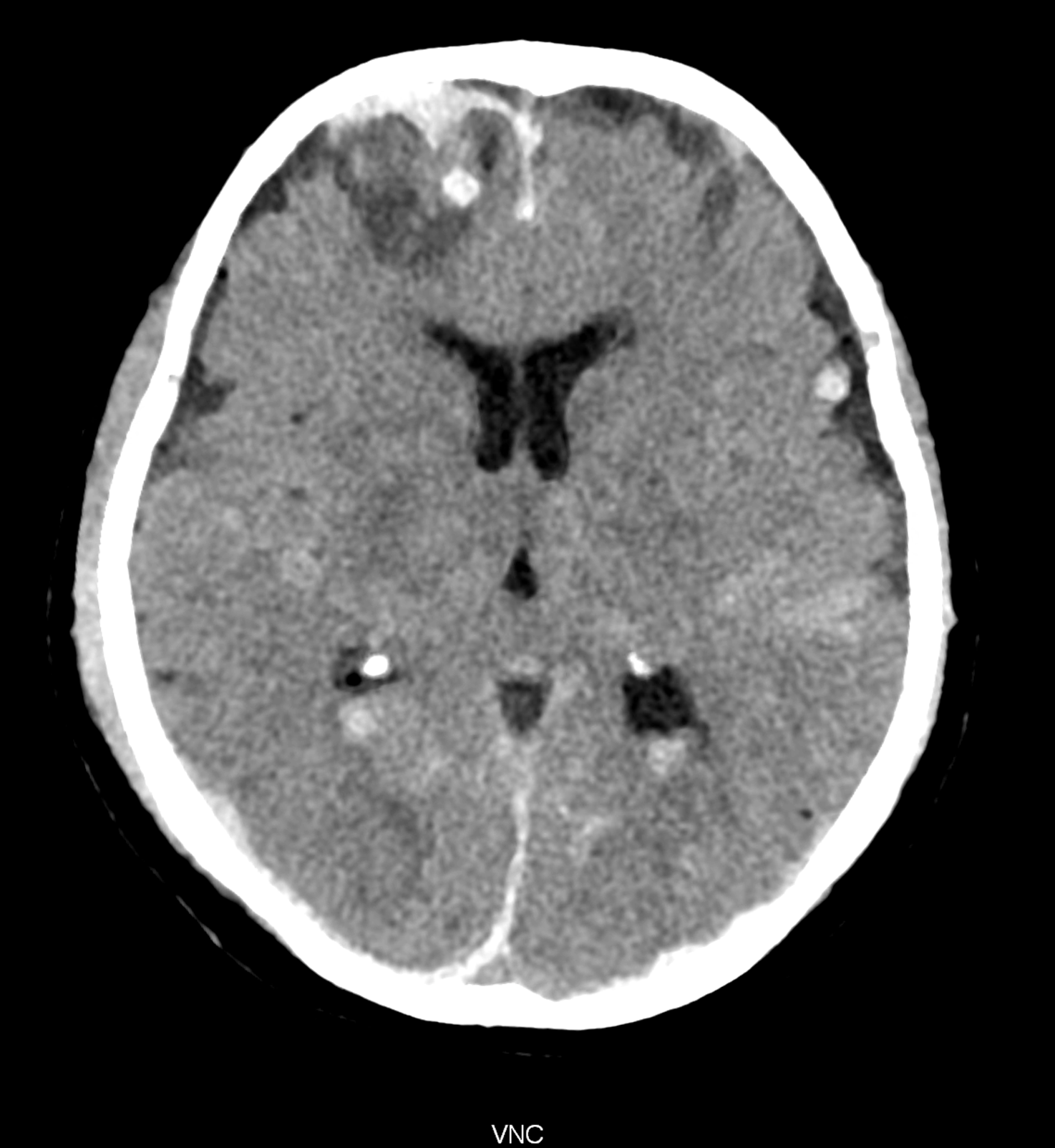
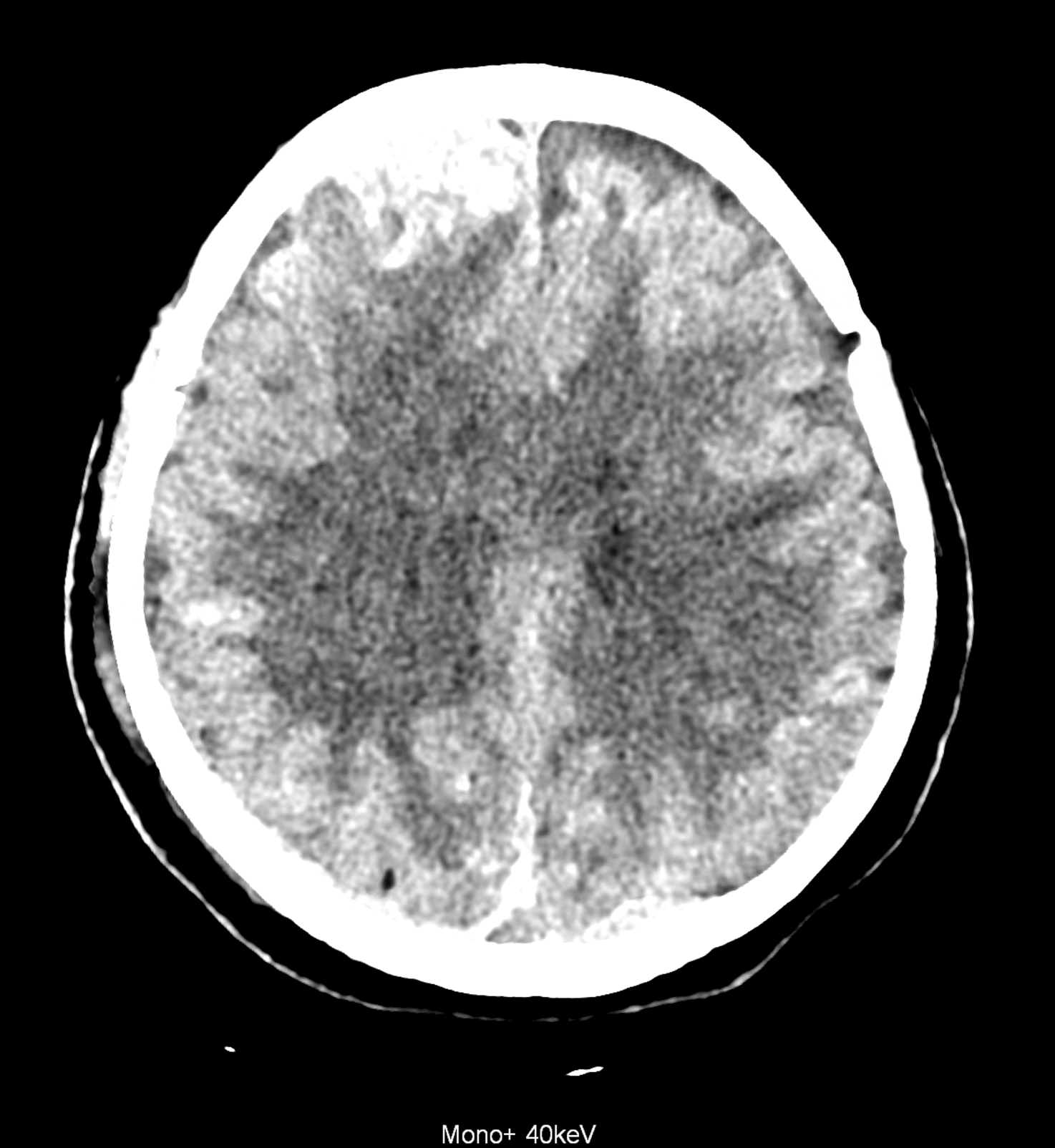
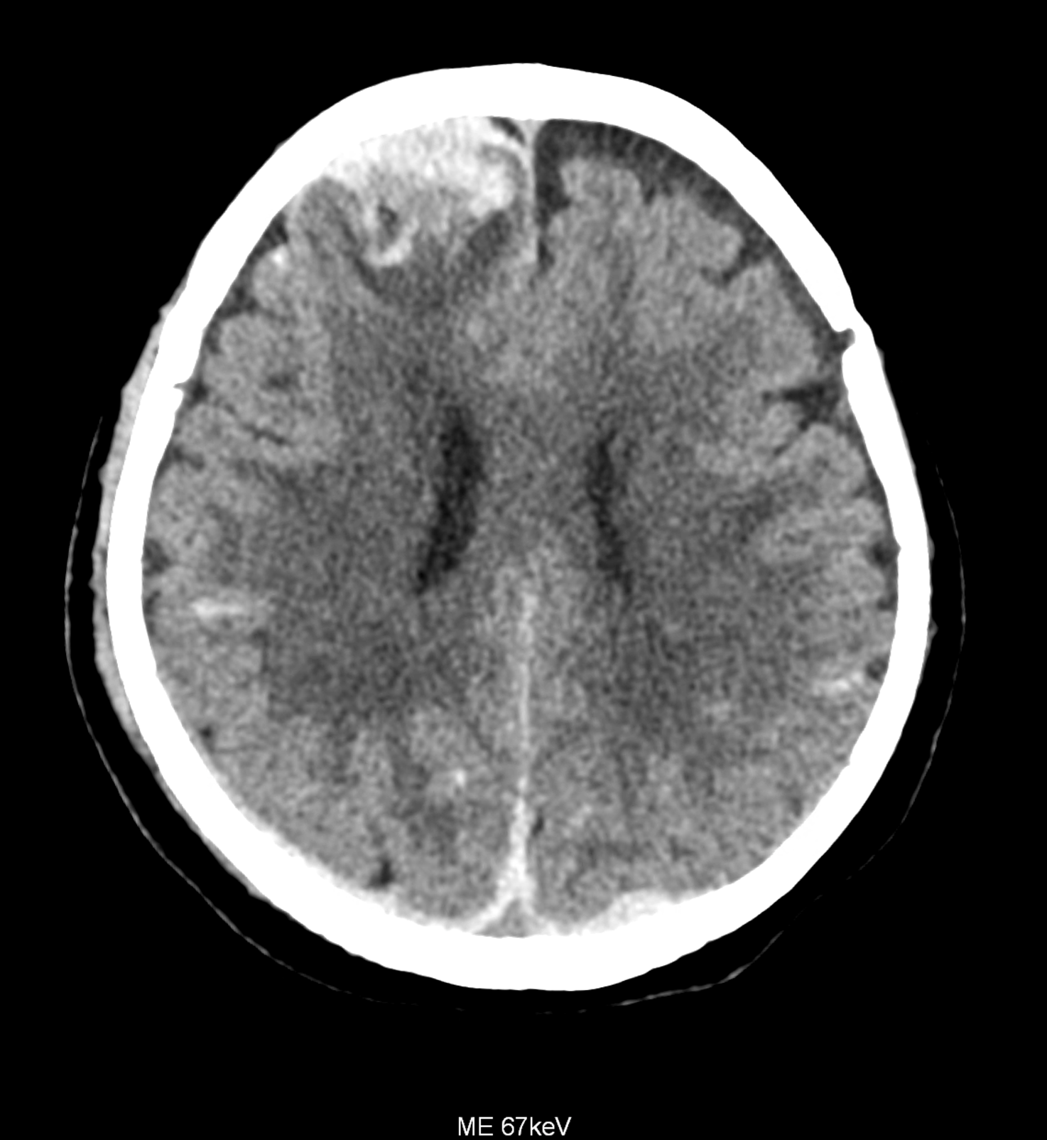
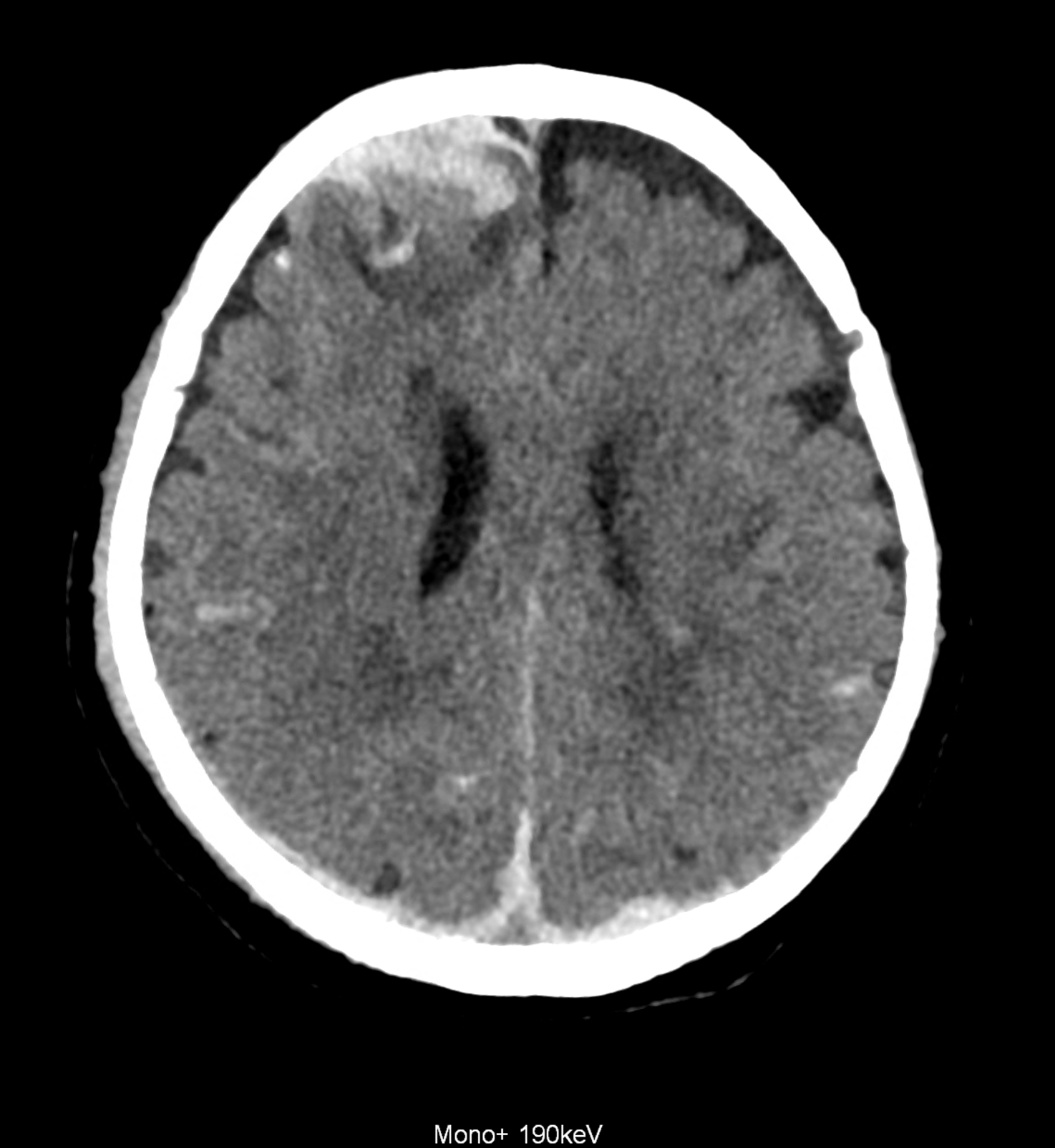
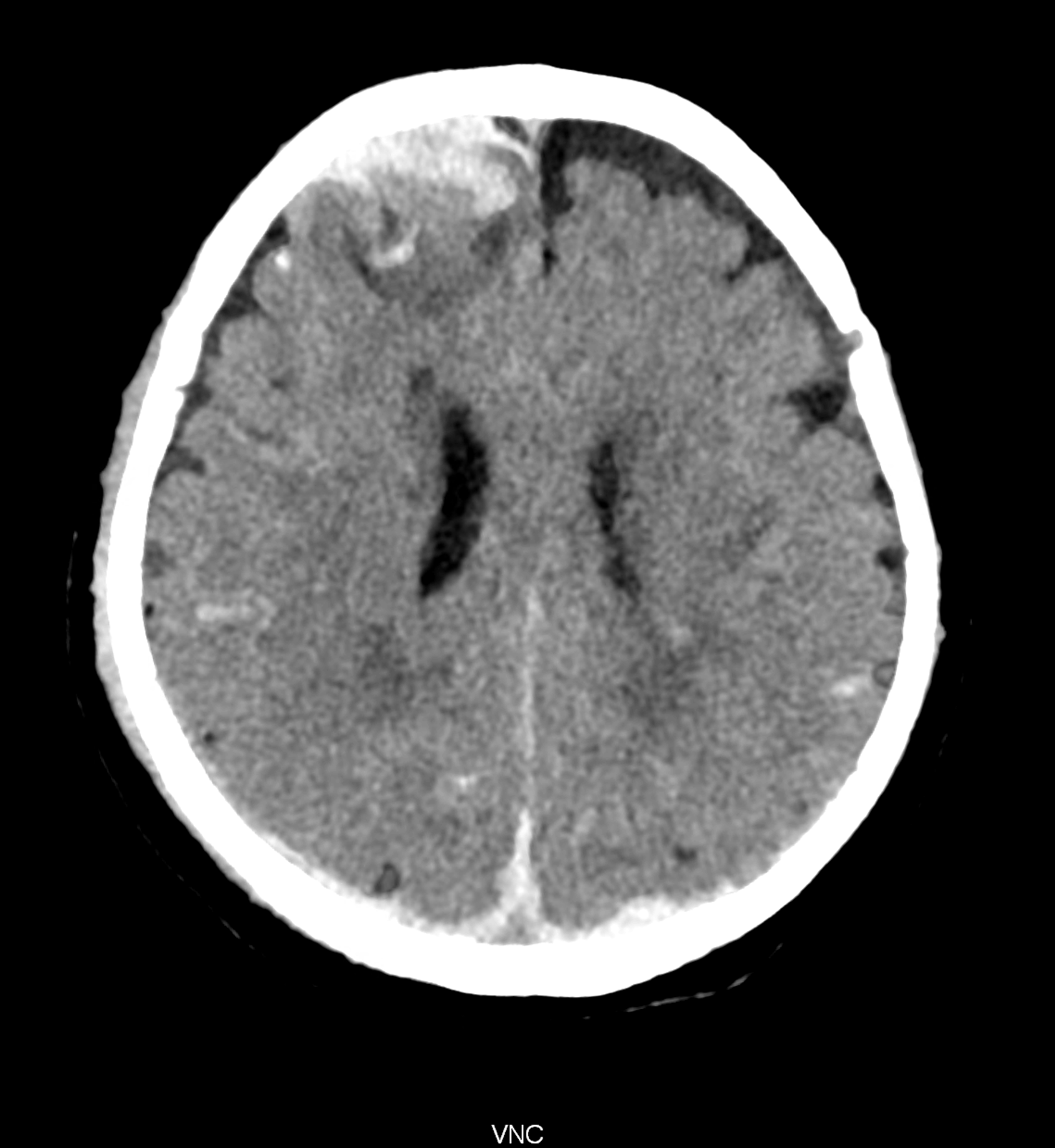
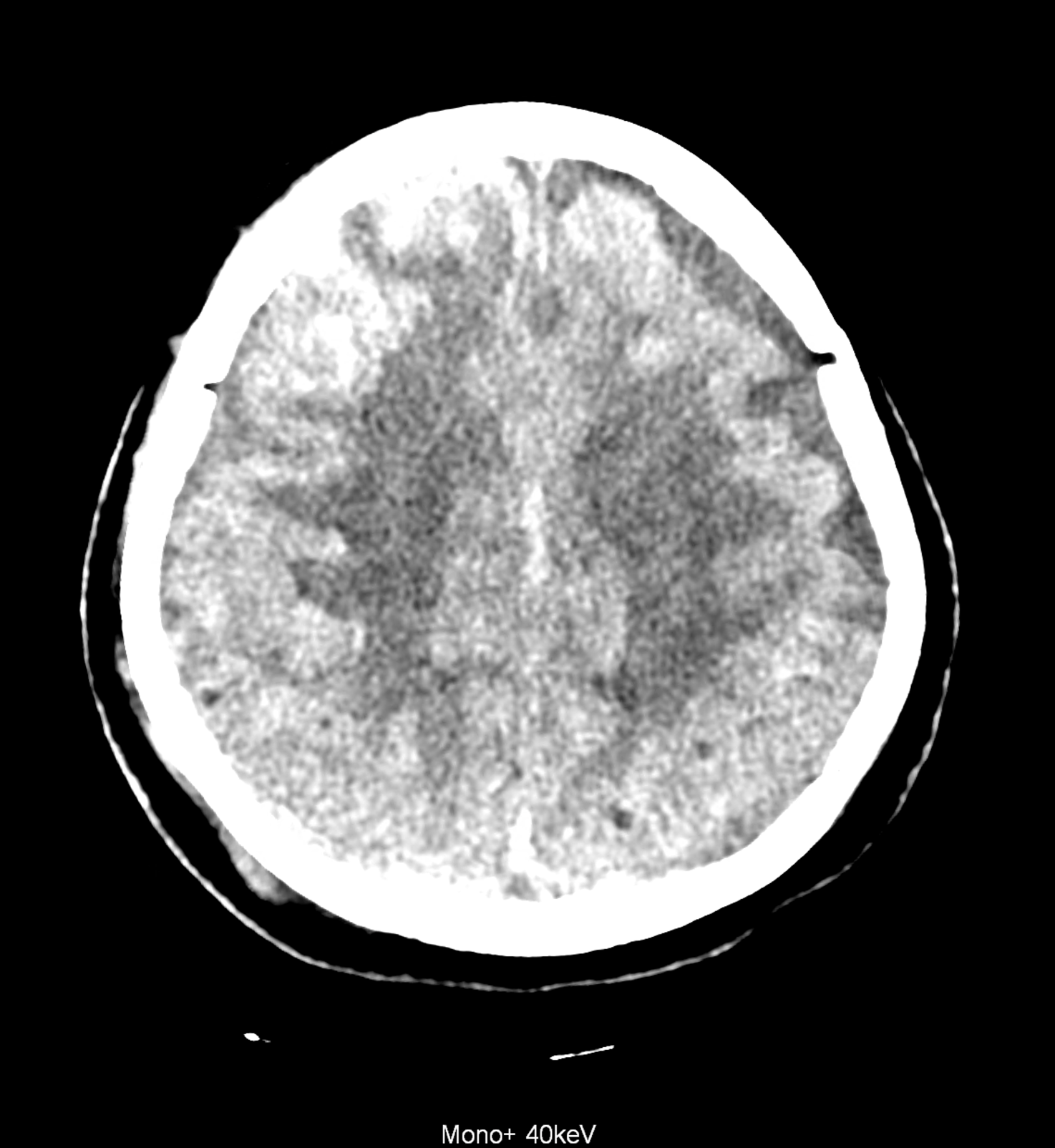
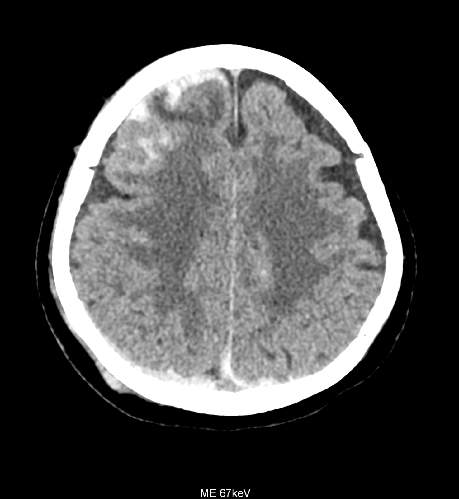
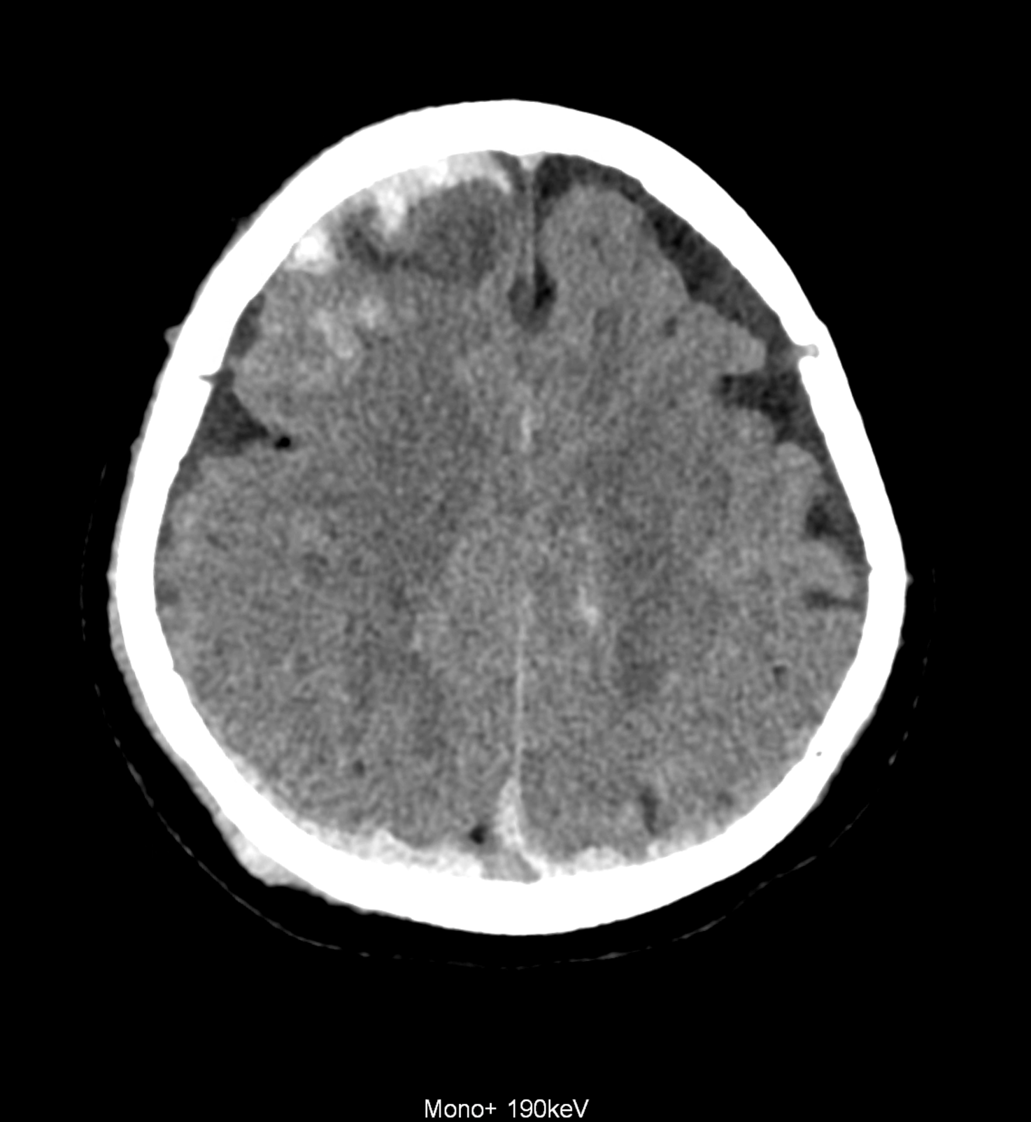
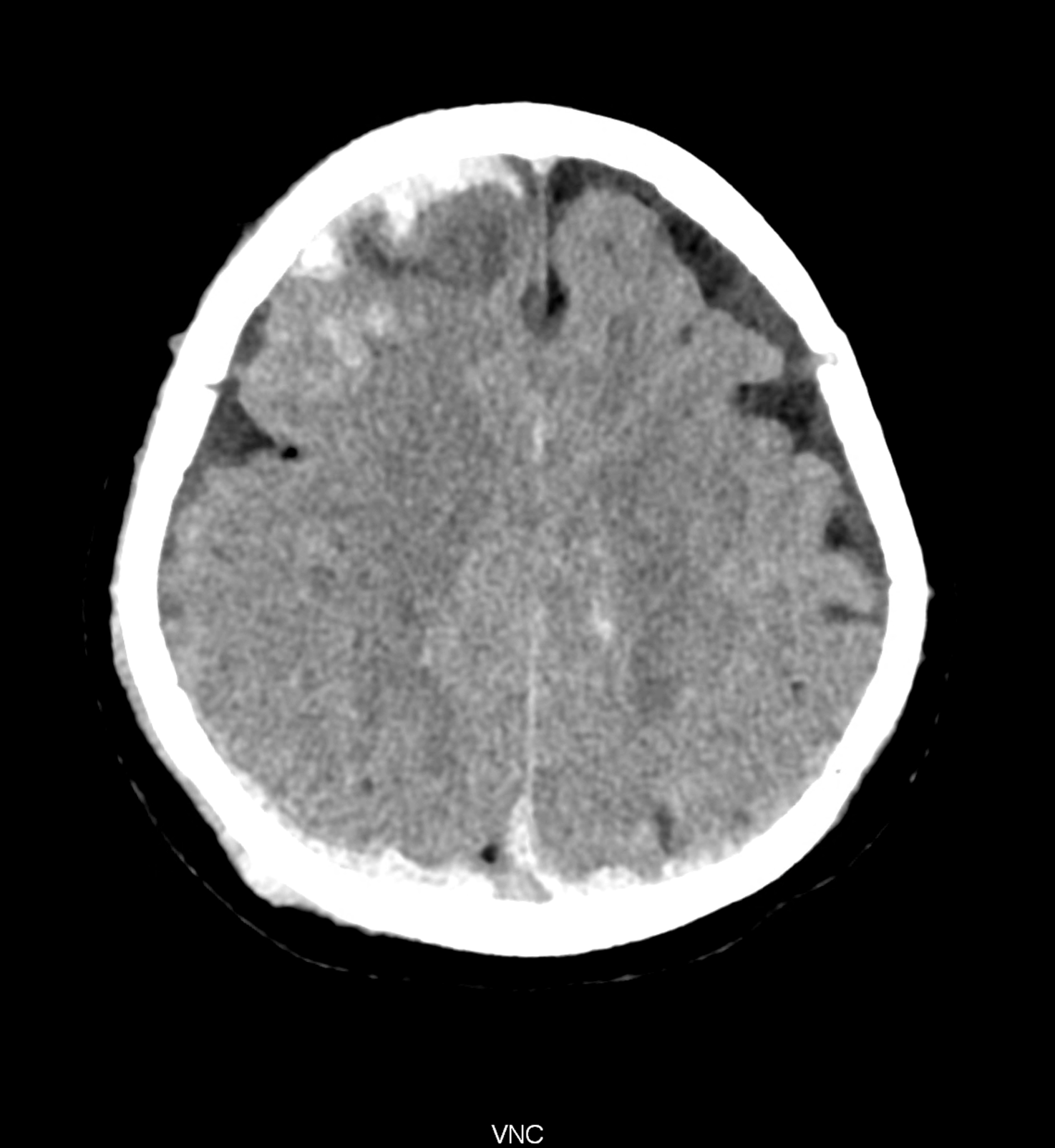
Bleeding is characterized by different absorption characteristics than healthy brain tissue (both gray and white matter) and cerebrospinal fluid. Due to the higher proportion of proteins with amino acids with disulfide bonds, the signal intensity increases at higher energies, making it possible to use monoenergetic imaging at energies above 140 keV, and/or virtual non-contrast. The hemorrhage then becomes more hyperdense, while the rest of the brain tissue acquires a uniform low signal. In addition to bleeding, areas of extracellular fluid of vasogenic origin are also imaged, i.e., in areas around contusion-type brain tissue injuries.
case
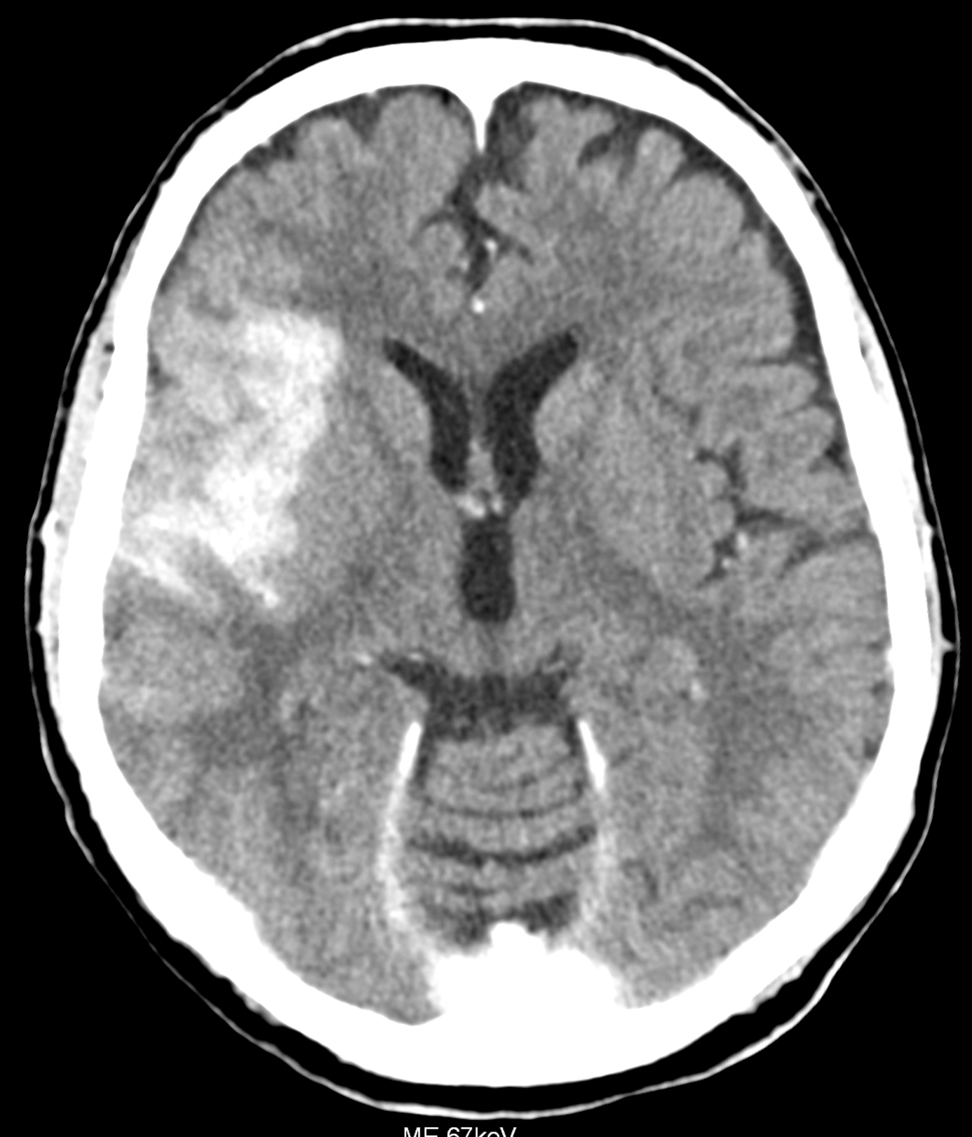
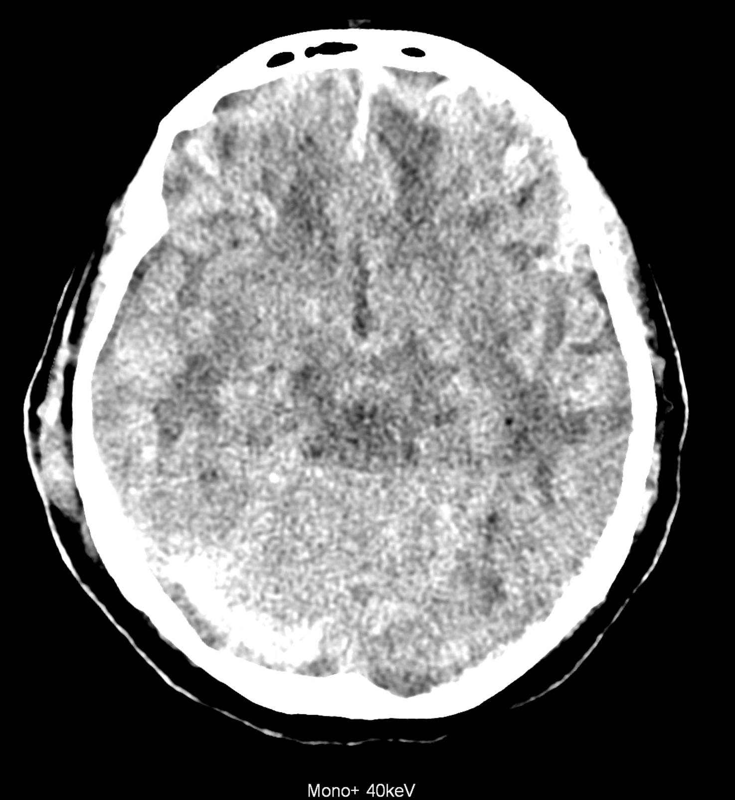
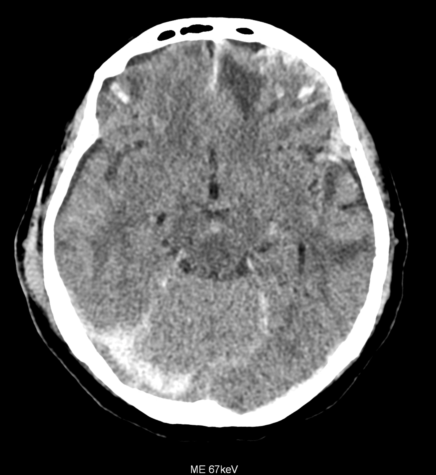
An example of the use of spectral imaging in complex brain injury, where contusions, diffuse axonal injury, subarachnoid hemorrhage, subdural hemorrhage, and blood in the ventricular system are present.
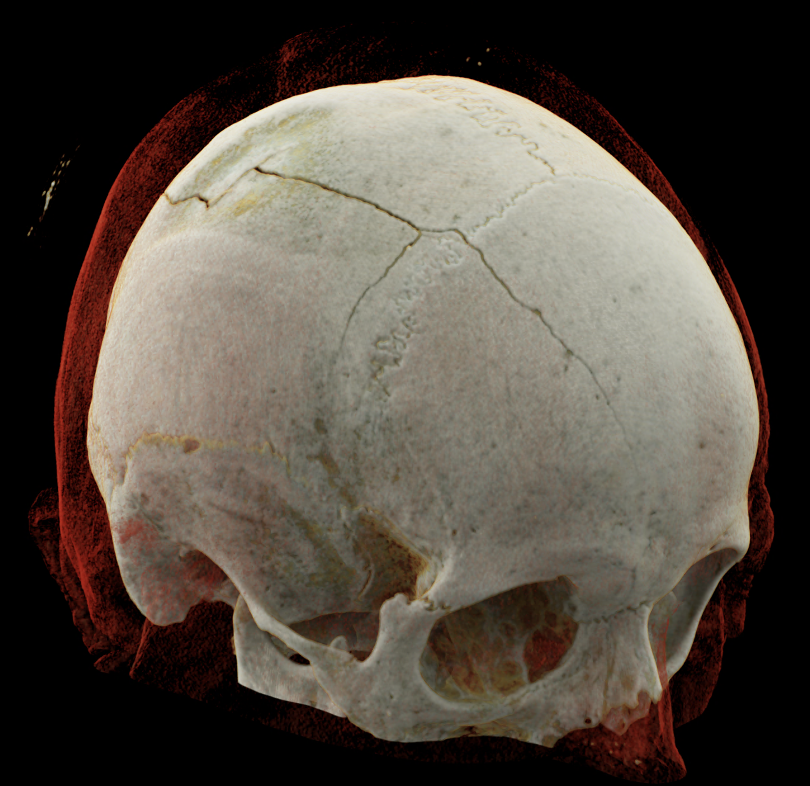
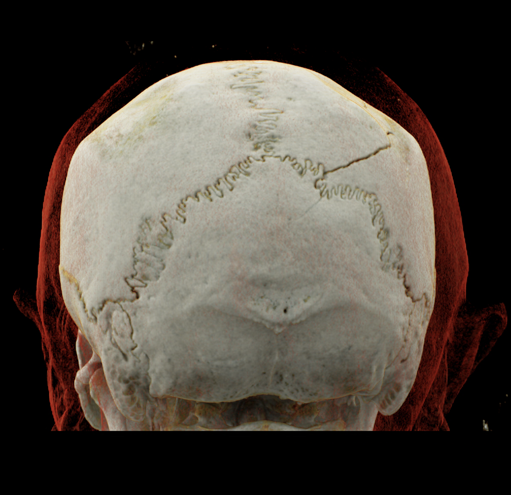
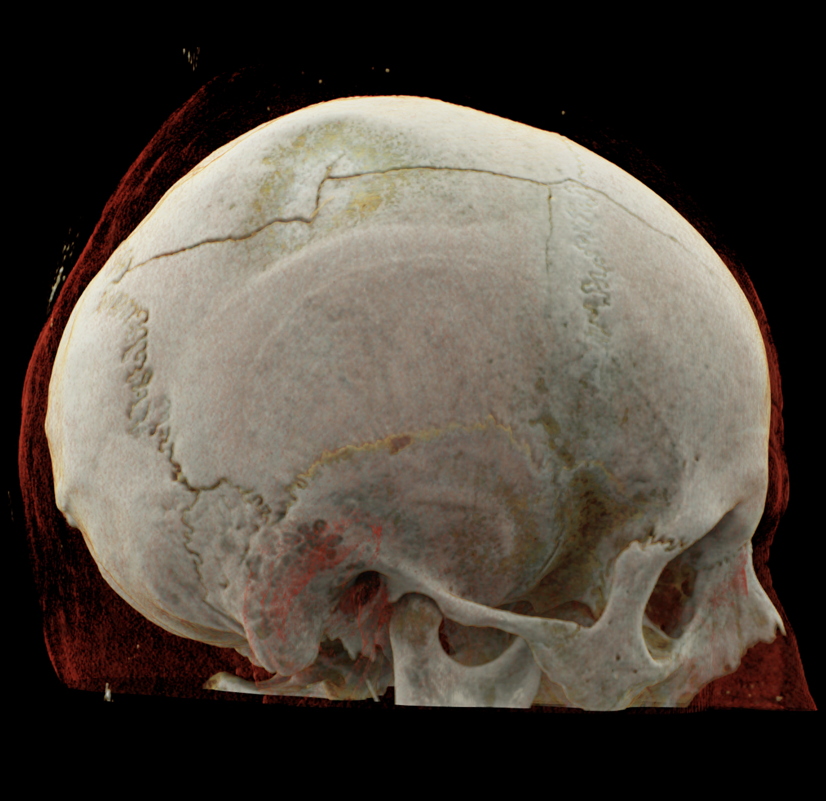
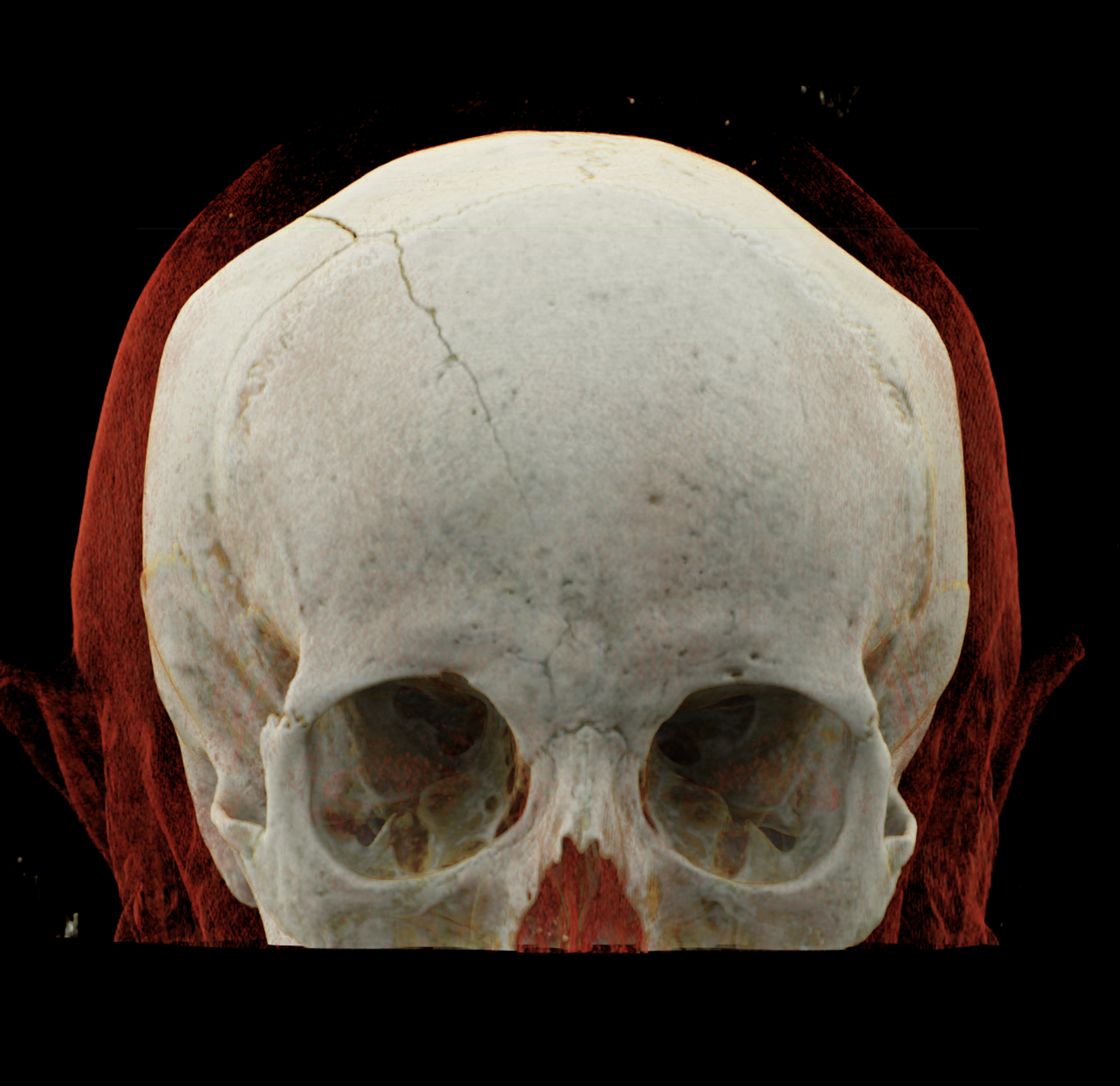
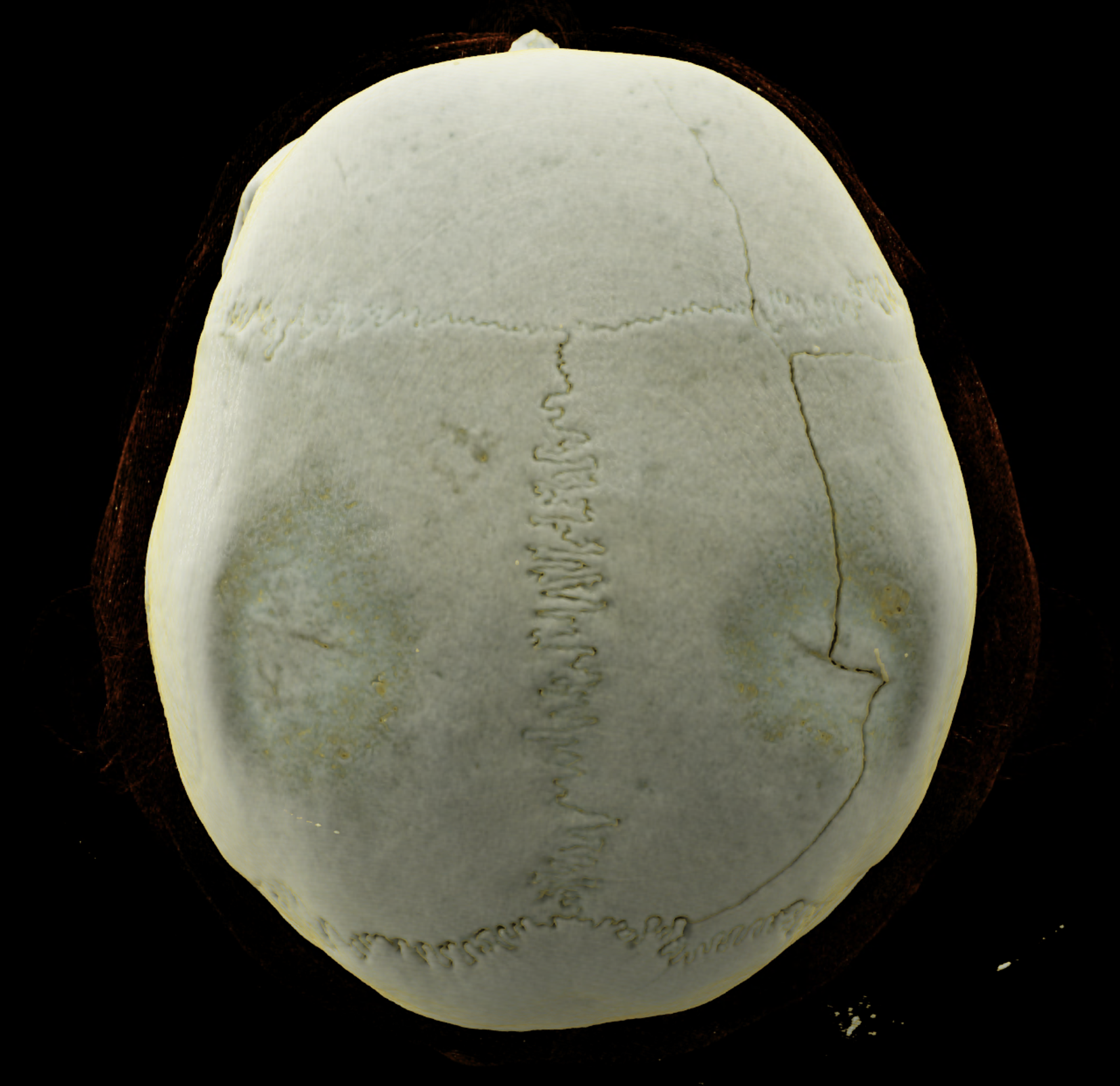
Three-dimensional reconstructions also show a complex skull fissure that extends from the frontal bone to the parietal and temporal bones.
Naeotom Alpha.Peak, University Hospital Pilsen, Czechia
comparison of the images with the energies of monoenergetic reconstructions 40 keV, 67 keV (standard conventional reconstruction), 190 keV and virtual non contrast (VNC)


volume rendered technique – display of the fissure course