ultra-high resolution imaging
the photon-counting detector is made from the small detector elements- respectively parts of the detector which are being the source of one single data trace. The smallest particular spot creating the data trace is the spot of 0.2 x 0.2 mm. These smaller detectors could be added together to create the complex data trace of square sized 0.4 x 0.4 mm. This detector element than could collect data with the inherent spectral information about he energy of quanta attenuated within the semiconductor material.
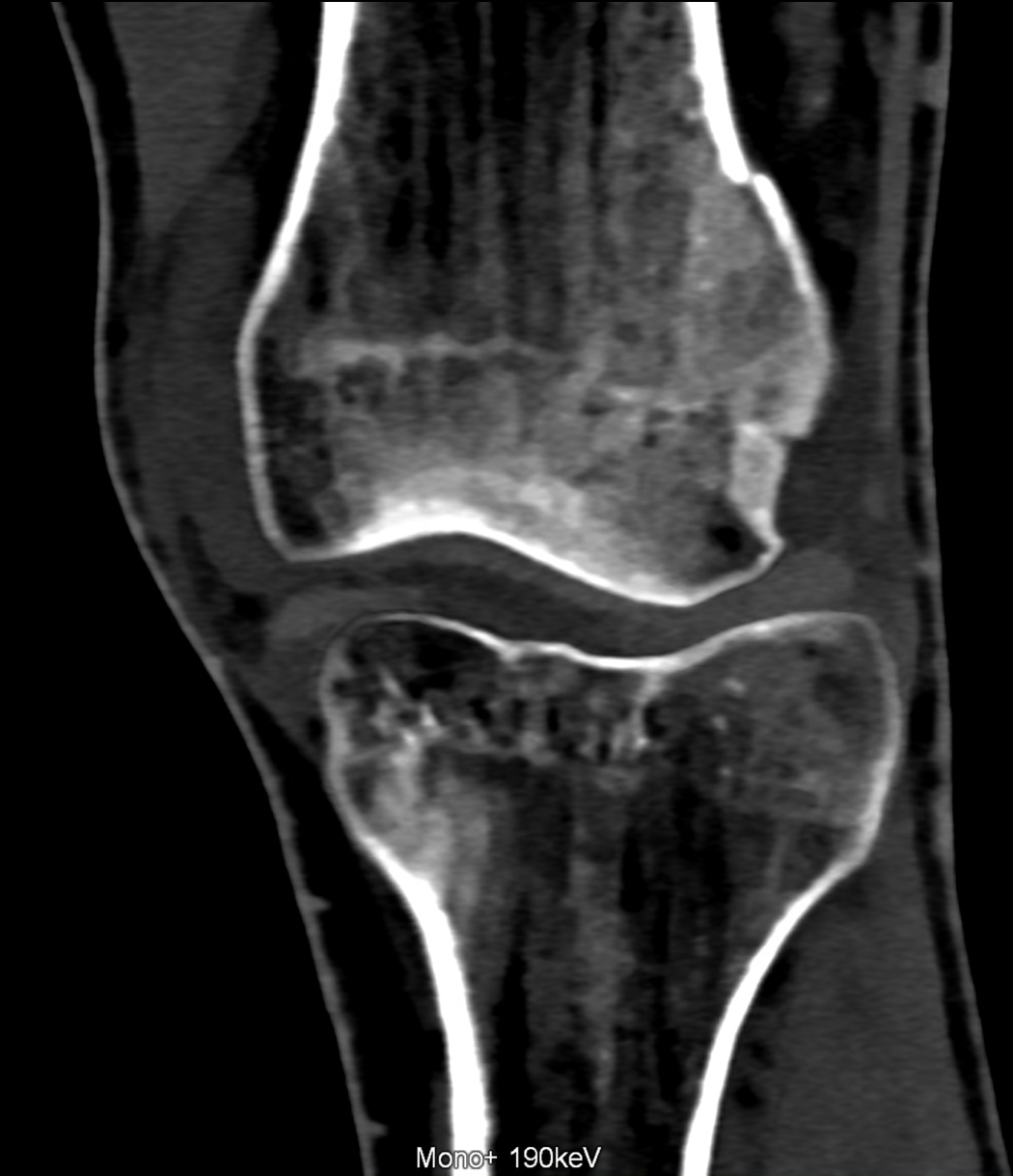
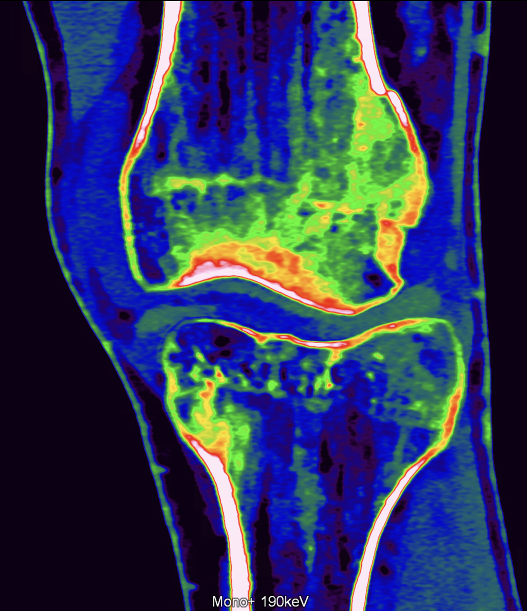
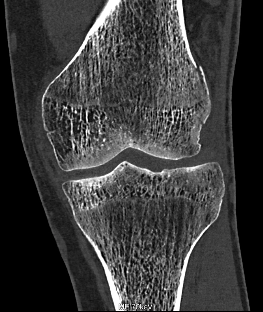
Photon-counting CT enables to use the smallest detector element with the size of 0.2 mm, the reconstruction with sharp kernels and the overlap of a half of the thickness improve the spatial resolution up to 0.1 mm, and in the same time the reconstruction of the data with inherent spectral information from the spatial voxels of 0.4 mm (made from four neighboring individual elements of 0.2 mm. Either the ultra-high spatial resolution , or spectral information derived from four neighboring elements could be used. Spectral acquired information enables the reconstruction of monoenergetic images in the range between 40 keV and 190 keV. The spectral imaging improves the detection of the edema on low-energy images and the intraspongious hematoma in high energy ones.
case report
19 years patient after ski-trauma with the abruption of the fragment form supracondylar portion of the femur. The distinct line of the fracture without dislocation is visible. The added information from spectral analysis shows the edema within the femoral condyle and also in the tibia condyle. The ultrahigh resolution image of the trabecular bone displays the irregularity of trabecular pattern caused the micro fractures inside the spongious bone of both condyles.
Naeotom Alpha.Peak, University Hospital Pilsen, Czechia
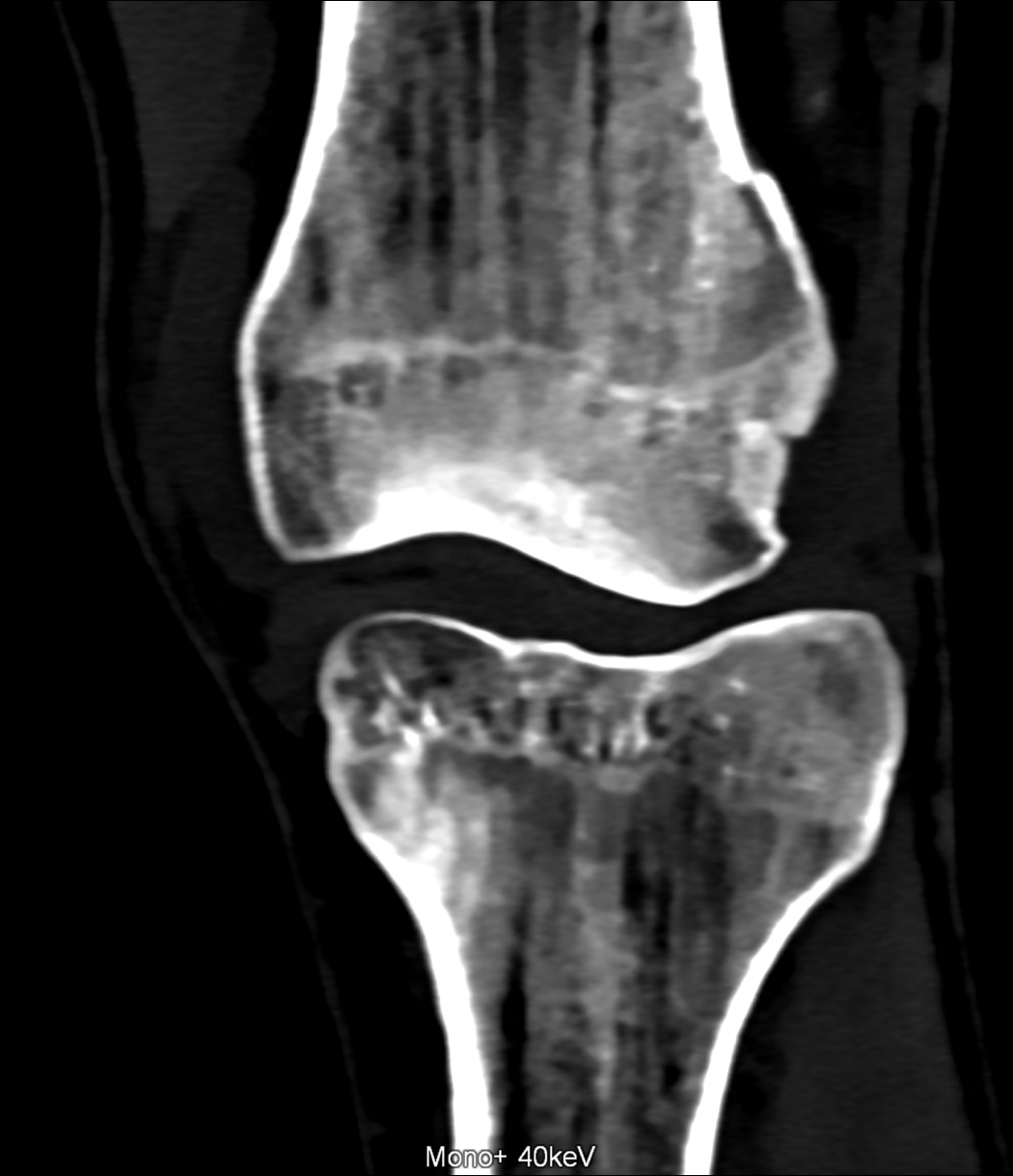
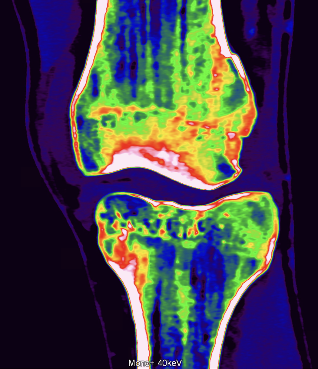
greyscaled and color coded monoenergetic images with 40keV (upper rows) and 190keV (lower)