Mycobacterium tuberculosis, like extremely aerobic microorganism, prefers the invasion into the best ventilated parts of the lungs. The chronic state of the post-primary tuberculosis occupies typically the apical segments of the upper or lower lobes. The cavities have quite regular inner surface and the relatively thick wall. The cavernous cavities have the communication with some larger bronchus. The bronchogenic spread is typical with the dissemination of the nodules along the bronchi, again mainly in upper lobes.
case report
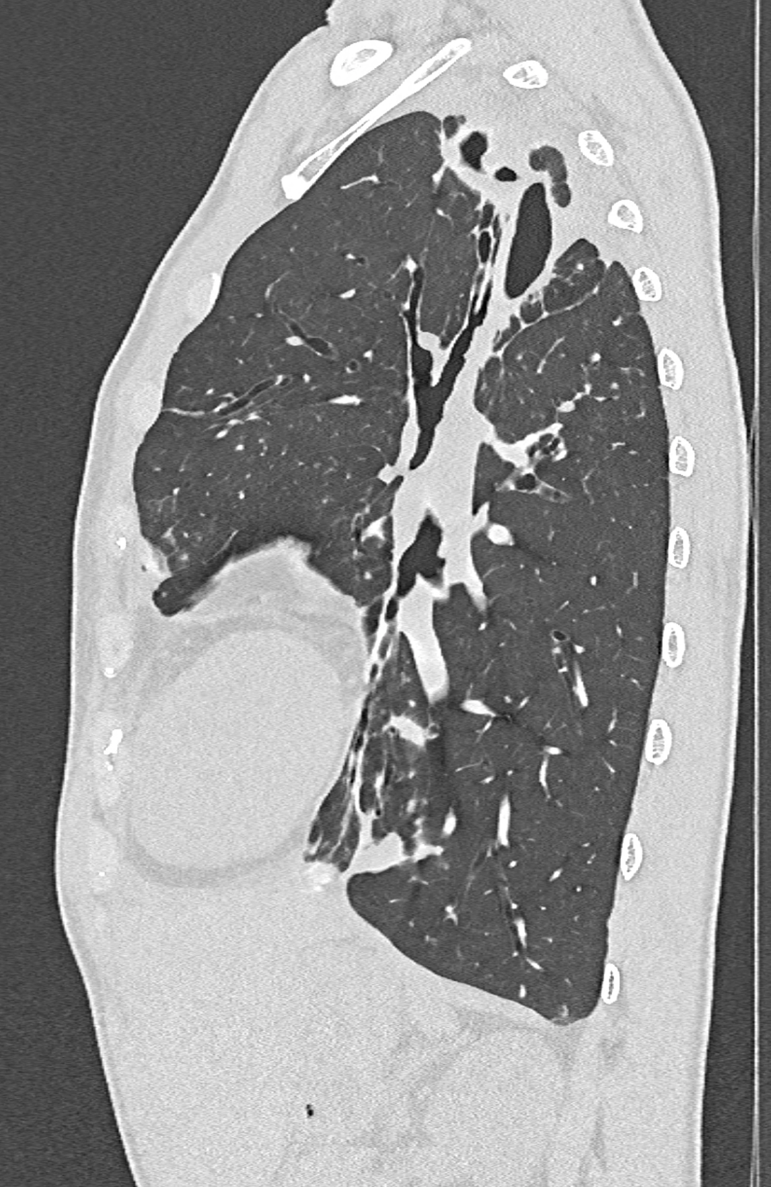
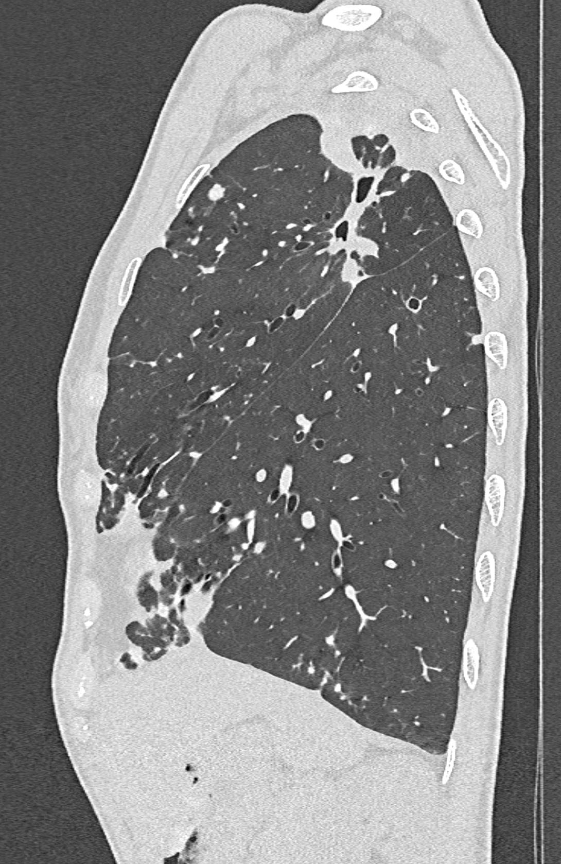
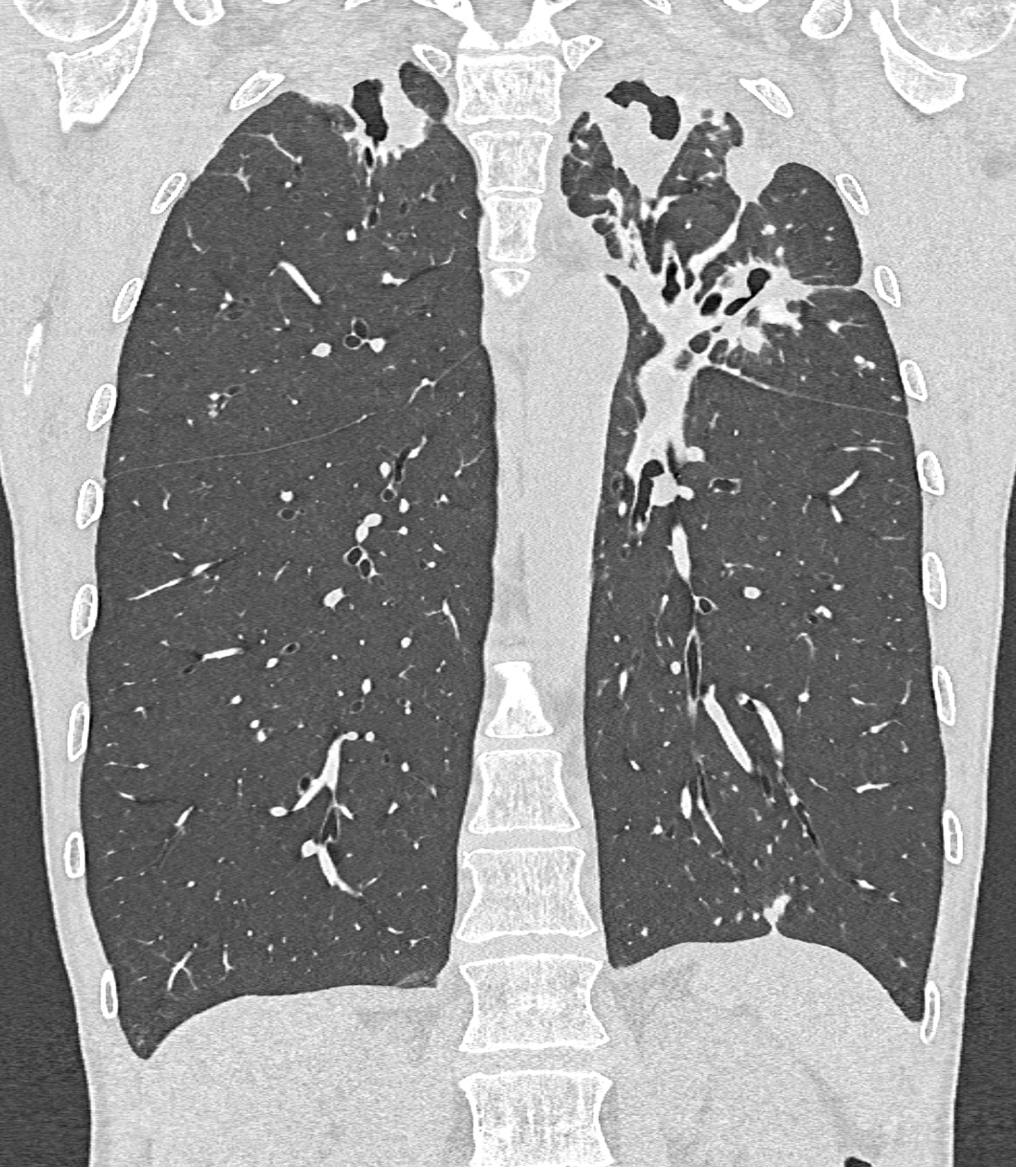
51 years old female with the six monthly history of microscopically and PCR confirmed Mycobacterium tuberculosis infection. The cavities are distributed mainly in the upper lobe of the left lung, where the communication with the B1/2 is present. The chronic course of the disease leads to the retraction of the hilar structures – the sign of the hilar cranialization. The active disease could be detected because of the distribution of smaller nodules along the bronchi in the anterior segment of the right upper lobe (S3). Ultra-high resolution using the detector element of 0.2 mm with the increment of 0.1 mm causes the resolution in cranio-caudal direction of 0.1 m. It improves the display of the communication of the bronchi and the detection of tiny nodules adjacent to the interlobular pleura.
Naeotom Alpha.Prime, University Hospital Pilsen, Czechia
the images document the cavernous cavities of both apical segments of upper lobes, note the distribution of the nodules in S3 at the axial images,