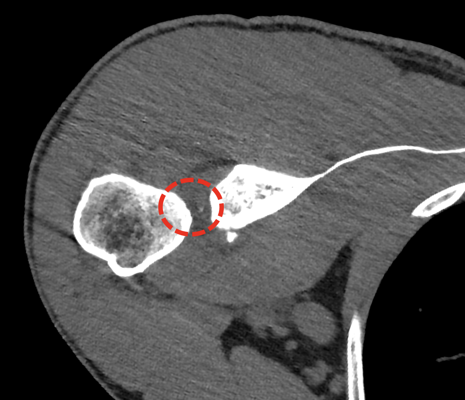
ultrahigh-resolution CT with the use of the detector element 0.2 mm with one half overlapping reconstruction resulted in voxel size of 0.1 m- Such a resolution enables to visualize very small fragment interposition between catlilaginous surfaces of the joints. The smallest possible imaging improves the detailed analysis of the position of the fragments.
case report
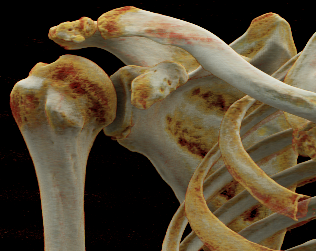
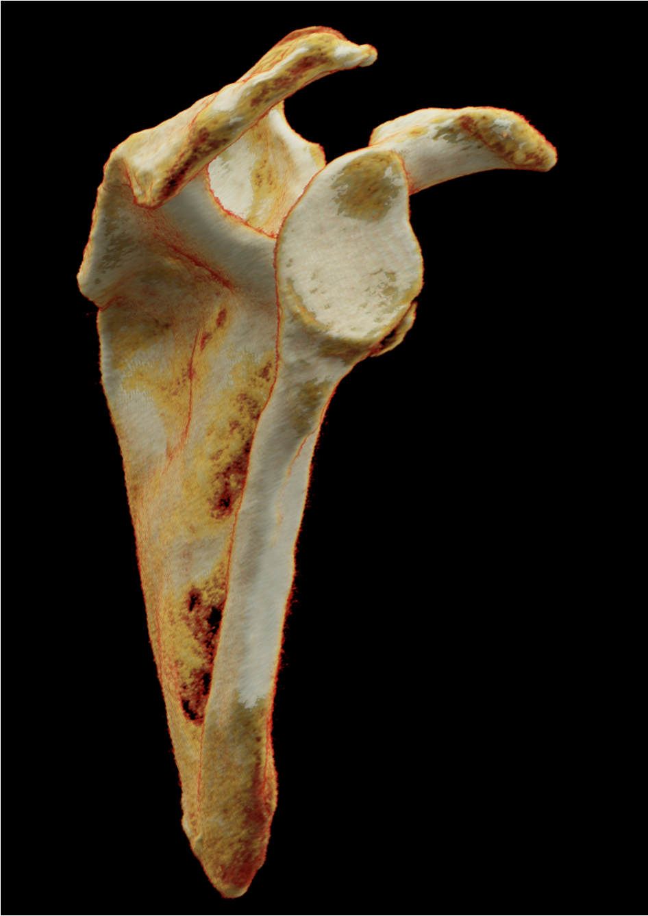
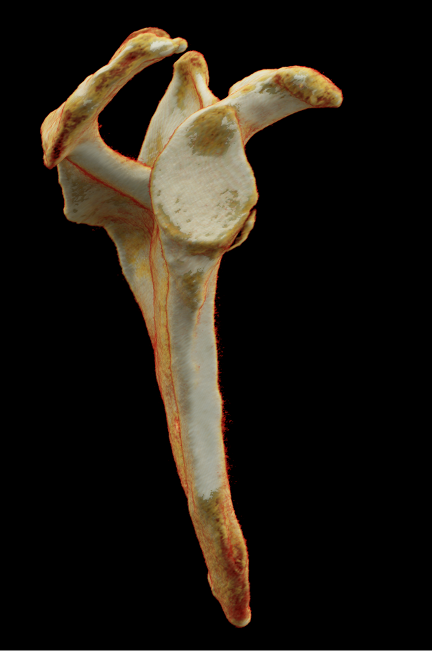
in middle aged male, it was made the reposition of the glenohumeral axillary luxation, the dislocation was caused after sport injury. The reposition was successful, the imaging was performed due to the suspected injury of the antero-inferior rim of the glenoid cavity. The abruption typical for Bankart osteochondra lesion is seen, but additional important finding is the interposition of the smallest osseous fragment into the inferior part of the articulation fissure.
Naeotom Alpha Peak, University Hospital Pilsen, Czechia