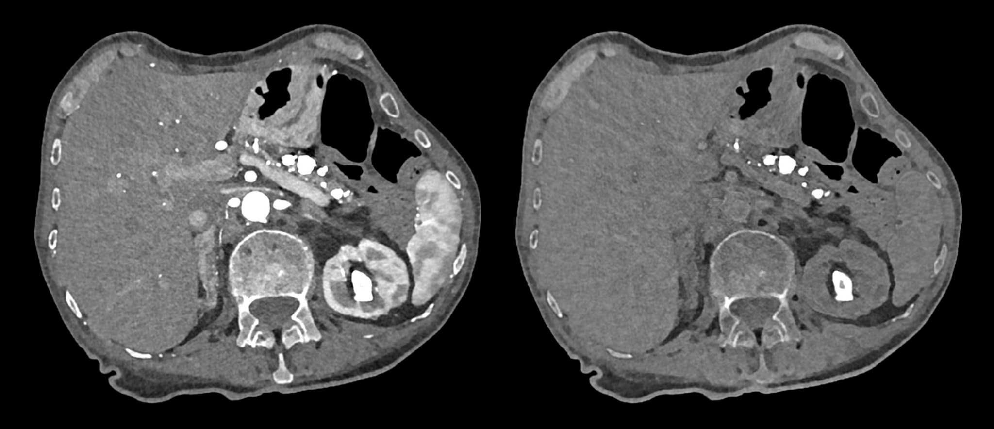
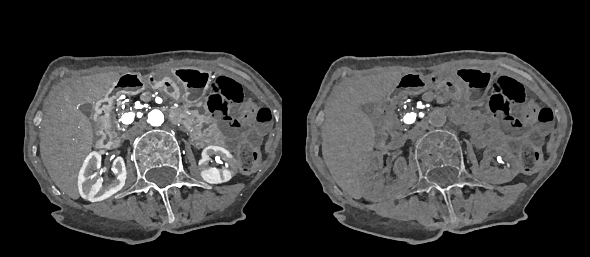
the spectral information of the CT images could be exploited in cases of the stones created in the severe hyperparathyroidism. The increased activity of the osteoclasts releases the phosphates and calcium ions from the bony mineral. The resulted hyperphosphatemia and hypercalcemia is the cause of the precipitation of the calcium in the pancreatic parenchyma and ducts, and/or in urinary outflow tract. The pancreatitis and renal colic could be consequent complications of hyperparathyroidism.
The imaging of the abdominal organs with the application of the iodinated contrast material shows the enhancement of the parenchymal organs. The visualization of the calcium containing stones could be improved with the suppression of the iodine related density using spectral application of virtual non-contrast images.
In 62 years patient with the confirmed hyperparathyroidism is seen the reticular structure of the vertebral bodies card by the activation of the osteoclastic resorption, this pattern is being called osteitis fibrous cystic Recklinghausen. The urinary stones are present in both kidneys. calcifications/stones are present in the pancreatic parenchyma and in the pancreatic duct. Irregularity of the renal cortex enhancement on the left shows confirms the presence of the pyelonephritis. Virtual non-contrast imaging surpasses the signal of the all other structures except those, which contain calcium.
Naeotom Alpha Pro, University Hospital Pilsen, Czechia