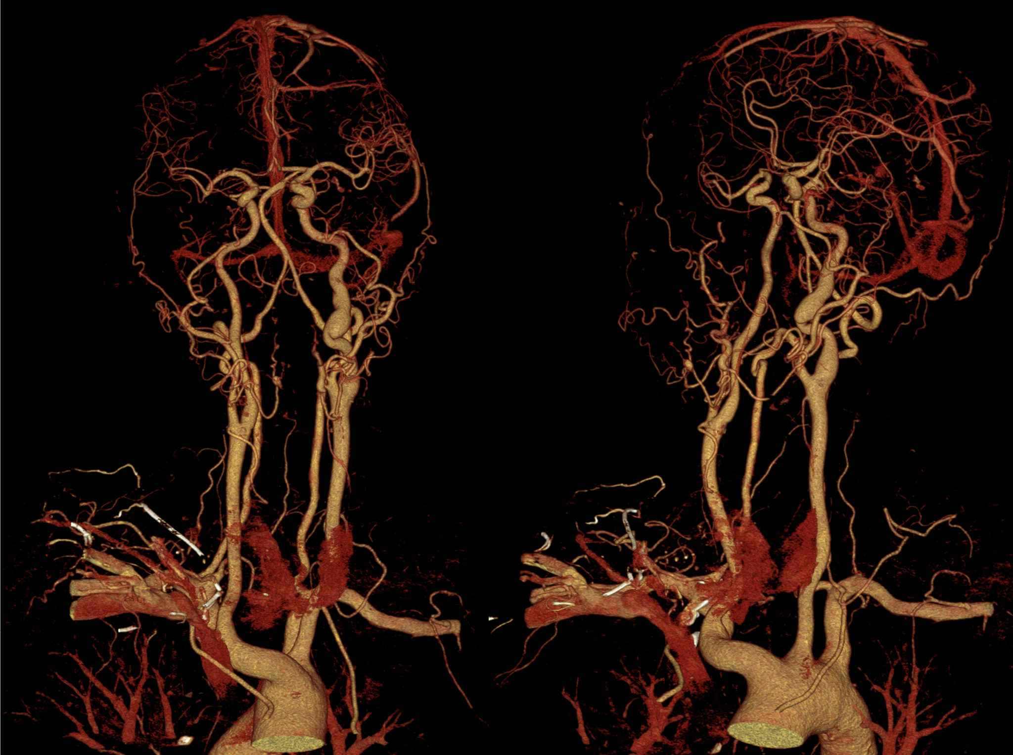
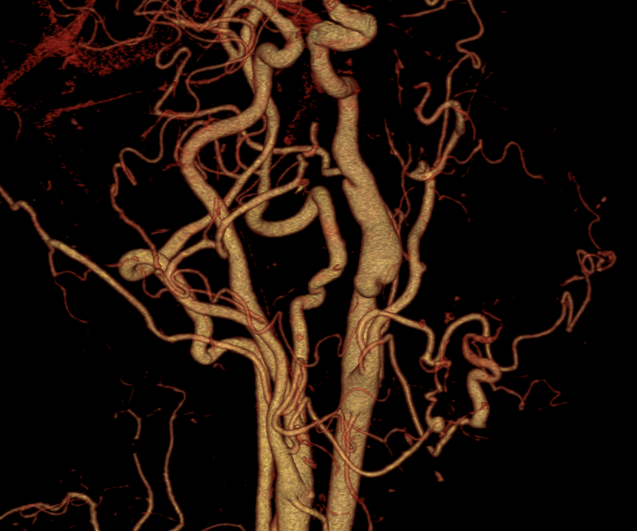
Fibromuscular dysplasia (FMD) is a rare blood vessel disorder in which some of the strong, flexible cells of arteries are replaced with cells that are more fibrous. Fibrous cells are less strong and also less flexible. Those changes in composition of the arteries leads to their becoming stiffer and more prone to damage. Typical localizations of the disorder are visceral arteries like renal or mesenteric, but very frequent is the involvement of the cervical arteries. This can lead to serious complications, including arterial narrowing (stenosis), weakening/bulging (aneurysm) or tearing (dissection) with consequent ischemia of the affected territories. At least 90 percent of adults with fibromuscular dysplasia are women. This disorder may be related to other connective tissue disorders, such as Marfan or Ehlers-Danlos syndromes.
Although the radiologic picture is variable, the typical CT finding is the irregularity of the lumen of the carotids, when some arteries are bearing aneurysmatic enlargement. Those aneurysmal enlargements could be multiple forming the typical sign of pearl necklace. In some individuals, the manifestation could be very weak like carotid web, or very serious with dissection, intramural hematoma and carotid occlusion. The most frequent localization on the neck level is the internal carotid.
Naeotom Alpha Peak, University Hospital Pilsen, Czechia
Comments are closed.