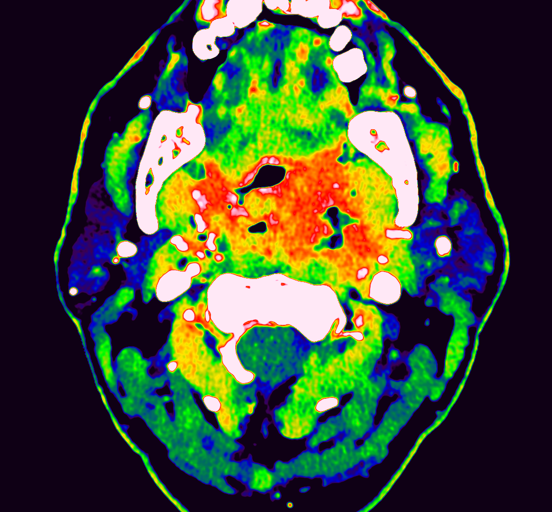
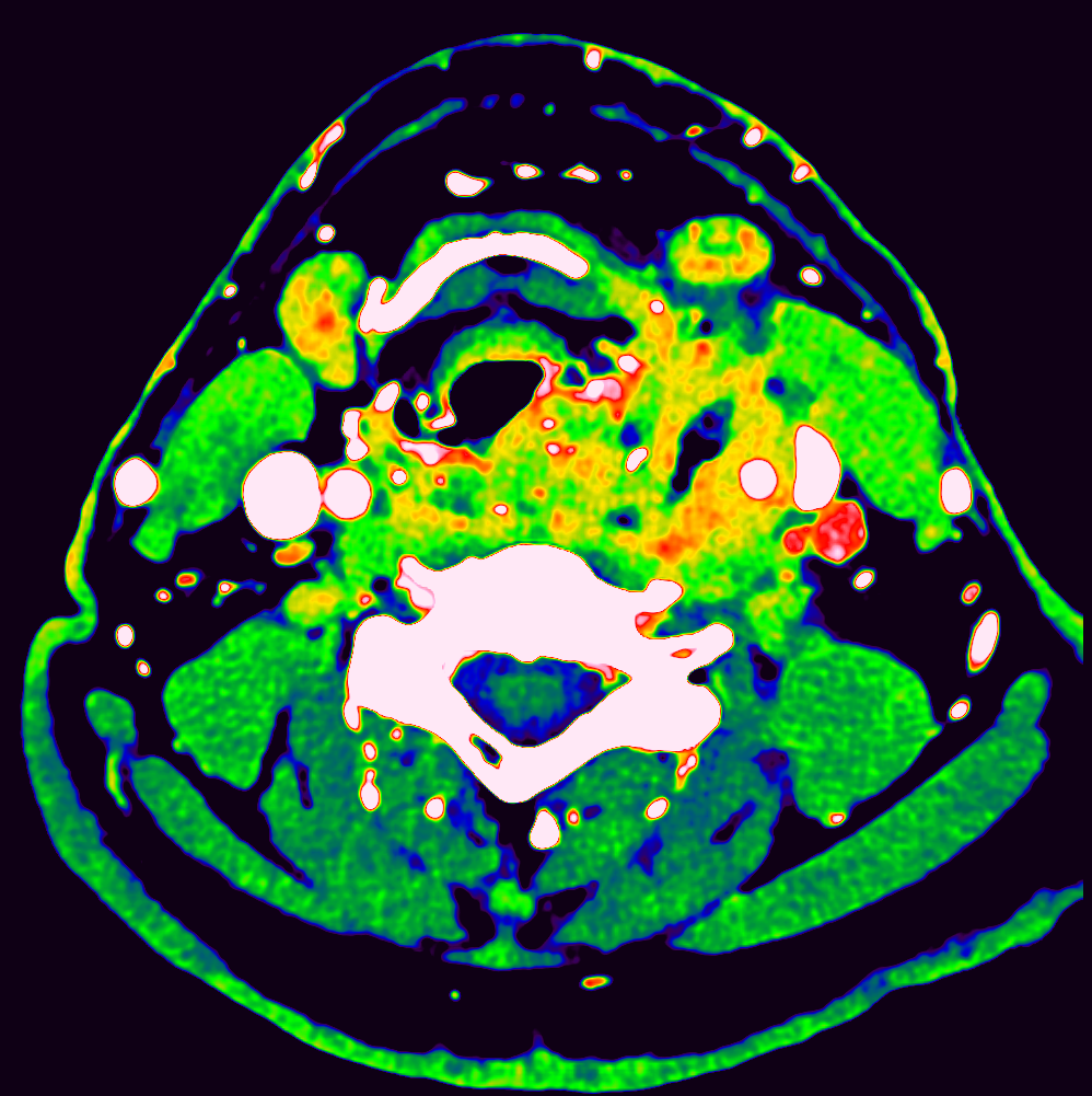
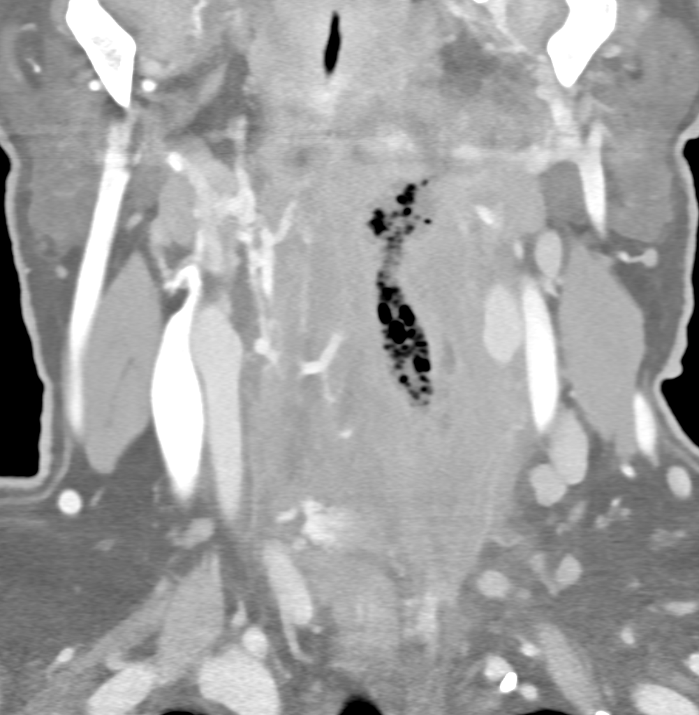
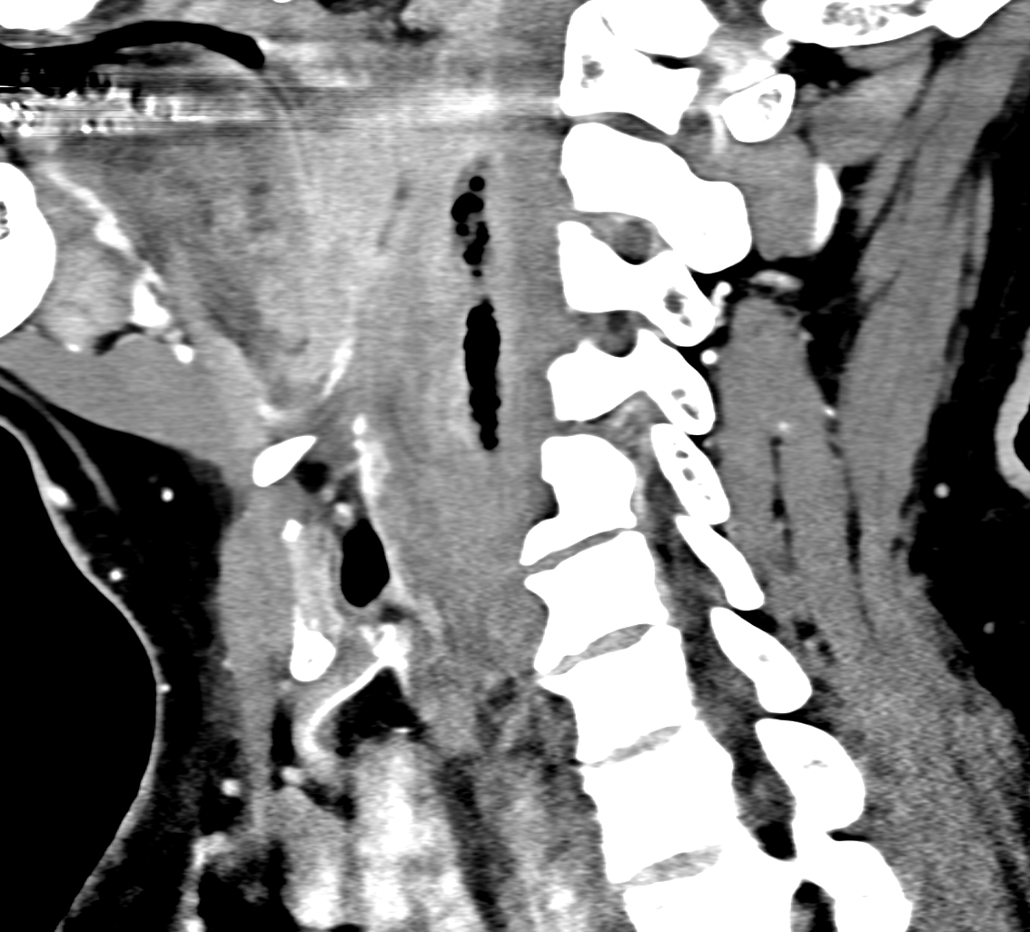
high-resolution imaging of the head and neck improves the detection of the spatial distribution of the edema, enhancement of the tissues, vascular structures. Phlegmonous infiltration with the gas inclusions in the dissecting spread of the infection from the paratonsillar space down to the lower deep neck spaces. The anaerobic infection exhibit typically low-level enhancement due to the hypovascularization and hypoxic environment. Spectral information coud be used in the assessment of the enhancement within the margins of the infiltration. The smallest voxel of 0.4 combined with smaller field-of-view increased the confidence of the imaging with photon-counting CT
Naeotom Alpha Prime, University Hospital Pilsen, Czechia


Comments are closed.