the smallest detector data trace of the photon-counting CT takes 0.2 x 0.2 mm. The reconstruction of 0.2 mm thin images with 0.1 mm increment with sharp kernel Bl64 enhances the resolution of the structures of the lung parenchyma. The resulted in-space resolution reachs 0.1 mm, to one third than the thickness of the human hair. The most important increase of the image quality is seen at the sagittal or coronary multiplanar reconstruction.
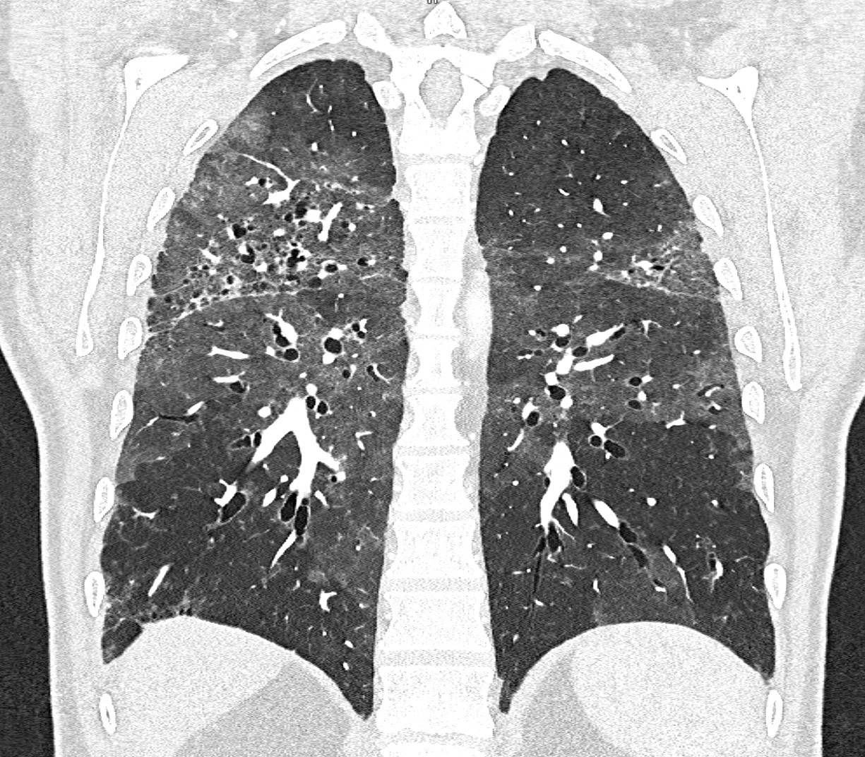
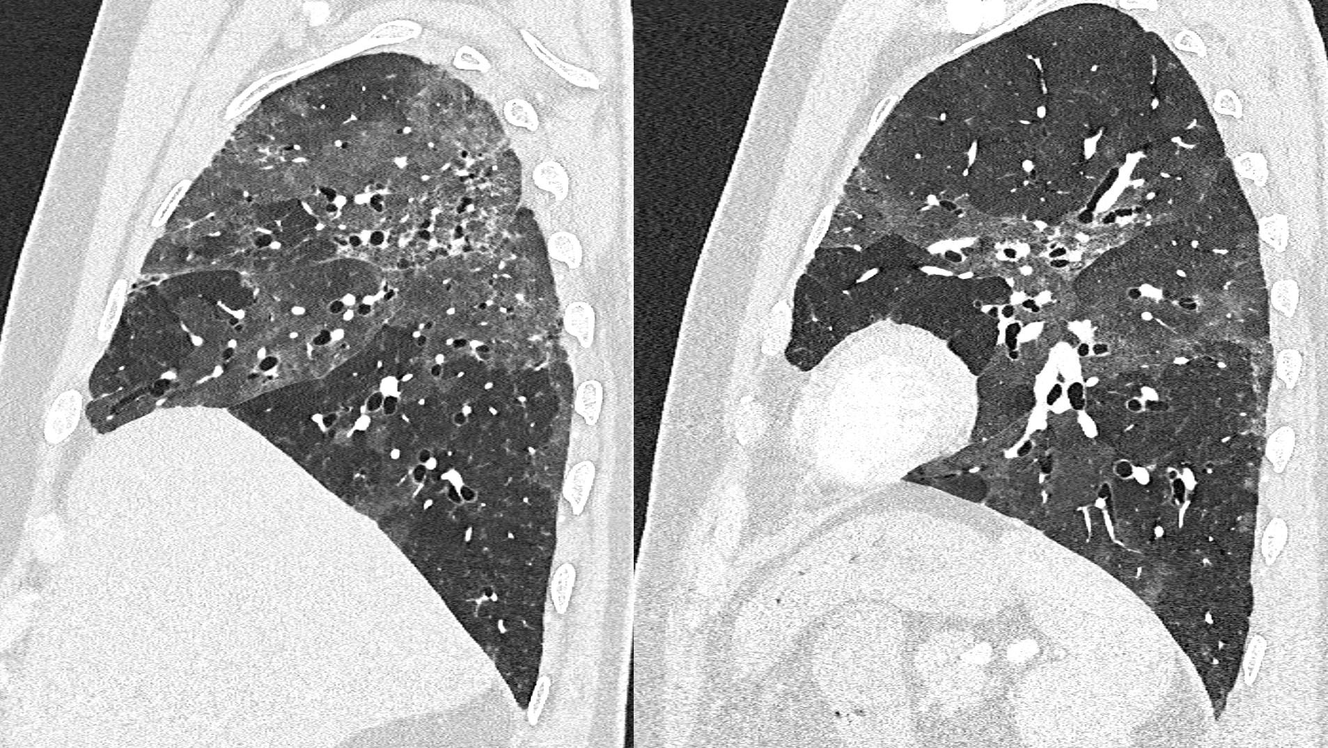
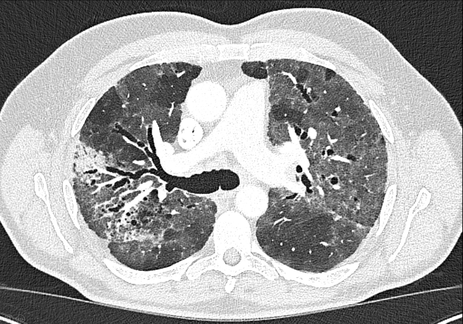
The images represent chronic hypersensitive pneumonia with advanced interstitial changes and traction bronchioloectasies
acquired with ultra-high-resolution mode QuantumPlus, Naeotom Alpha Prime, University Hospital Pilsen, Czechia
Leave a Reply